
Transmission or projection images use a radioactive source placed behind the target to project radiation through the target. The structures of the target cast shadows on the other side, where the image is then taken.
The most common form of projection image is the everyday X-ray (more properly referred to as a radiograph). While medical imaging is often thought to be a hot, exciting, and rapidly progressing field, 90% of all medical images taken every day are simple X-rays.
|
| an example of an X-ray. |
By taking a lot of X-ray images at a range of angles around the body, one can create a three-dimensional reconstruction of the anatomy. This process is known as Computed Tomography or CT (also known as Computed Axial Tomography or CAT scans--the "axial" and the "A" are often left off these days).
 |  |
| brain | thorax |
| CT image examples | |
Notice that the image slices across the axis of the body instead of projecting through it.
Ultrasound is a form of projection imaging using high-frequency sound waves, but rather than seeing shadows, it sees reflections (echoes).

Emission imaging involves putting the radiactive source inside the target. The radiation emitted by the target is directly imaged. Actually, not quite directly--as with projection imaging the anatomy of the patient casts shadows as the radiation leaves the body.
While the important information in projection imaging is the anatomy (the shadows cast by the body), the important information in emission imaging is the distribution of the radiation source within the body. These radioactive isotopes are usually bound with specific pharmaceuticals that distribute themselves in certain ways according to body function.

| Example of an emission image from nuclear medical imaging. The noise is typical of nuclear medical images. |
Common forms of nuclear medical imaging are photon-emission tomography PET and single-photon emission tomography SPECT.
A recent form of emission imaging is Magnetic Resonance Imaging or MRI, which causes certain molecules of the body to resonate and give off low-level radiation in the FM radio range. This signal can be detected outside of the body and the distribution of the resonating molecules can be formed into an image. The most common form of MRI involves imaging single-proton nucleii in the body (i.e., hydrogen, which appears in large quantities in the body in the form of water). This makes it more sensitive to soft tissues than X-ray imaging.
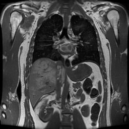 |
| MRI image example |
Since different images capture different features (CT capturing hard structure, MRI capturing soft tissues, PET/SPECT capturing physiological function, etc.) one can combine images from different modalities to give a more complete picture. This requires aligning the multiple images so that they correspond spatially--a process known as image registration.
One of the most interesting applications of multimodality imaging is the Visible Human Project conducted by the National Institutes of Health. This project is imaging a cadaver with MRI, CT, and cryosection imaging.

While multimodality imaging is common in medical imaging, it is also often used in other areas as well.
Maintained by John Loomis, last updated Jan 1, 2003